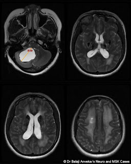
Clinically middle age male with history of progressive headache for last 5months.
CT brain shows multiple dense bilateral basal ganglia calcification.
DDs thought were like Fahrs disease, hypo thyroidism, hypo, pseudo hypo para thyroidism.
However patients thyroid and para thyroid profile was normal and the posterior cranial fossa cyst could not be explained even by Fahrs. With a possibility of a concurrent posterior fossa tumor, patient was referred for MRI brain with contrast.
MRI brain with contrast showed dense calcification in bilateral basal ganglia. The posterior fossa cyst with debris level. No abnormal enhancement along wall of cyst or enhancing eccentric solid nodule depicted on post contrast MRI which ruled out tumor nodule.
The bilateral symmetric confluent T2 white matter hyper intensity which represents an associated leukoencephalopathy.
Imaging wise primary diagnosis given was LCC.
Due to mass effect, fourth ventricle compression by the posterior fossa cyst leading to obstructive hydrocephalus, patient underwent posterior fossa craniotomy and excision of posterior fossa cyst was done. Histopathology revealed nonspecific cyst without any tumor cells keeping with MRI diagnosis of LCC.
LCC or Labrune syndrome syndrome MRI
Leukoencephalopathy, brain calcifications, and cysts (LCC) also known as Labrune syndrome, an extremely rare with only near 10 cases reported so far in the medical literature.
The condition is caused by homozygous or compound heterozygous mutations in the SNORD118 gene on chromosome 17p13. Clinical presentation varies from spasticity, dystonia, seizures, and cognitive decline.
Etiopathogenesis of LCC is still a matter of debate. Obliterative microangiopathy has been found on histopathological examination as the basic abnormality, the cyst formation is due to necrotic process secondary to obliterative microangiopathy and calcifications is dystrophic in nature. White matter changes result from changes in water content rather than abnormality of myelination.
Another entity which deserves a special mention is cerebro retinal microangiopathy with calcifications and cysts is a distinct genetic disorder due to CTC1 gene problem. Similar leukoencephalopathy, cysts, and calcification have been reported in few cases in association with Coat's disease, described as "Coat's plus. Coat's disease is unilateral retinal telangiectasia with exudation commonly occurring in boys sporadically without systemic features. However, in Coat's plus, there is bilateral retinal telangiectasia with exudation along with systemic features in the form of LCC.
In my case patient did not have any visual issues so that rules out retinal abnormality and the possibility of cerebro retinal microangiopathy.


Δεν υπάρχουν σχόλια:
Δημοσίευση σχολίου
Medicine by Alexandros G. Sfakianakis,Anapafseos 5 Agios Nikolaos 72100 Crete Greece,00302841026182,00306932607174,alsfakia@gmail.com,