Αρχειοθήκη ιστολογίου

Πέμπτη 10 Δεκεμβρίου 2020

Cadherins, Selectins, and Integrins in CAM-DR in Leukemia

pay.thelady61 shared this article with you from Inoreader

592733_Thumb_400.jpg

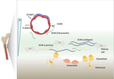
The interaction between leukemia cells and the bone microenvironment is known to provide drug resistance in leukemia cells. This phenomenon, called cell adhesion-mediated drug resistance (CAM-DR), has been demonstrated in many subsets of leukemia including B- and T-acute lymphoblastic leukemia (B- and T-ALL) and acute myeloid leukemia (AML). Cell adhesion molecules (CAMs) are surface molecules that allow cell–cell or cell–extracellular matrix (ECM) adhesion. CAMs not only recognize ligands for binding but also initiate the intracellular signaling pathways that are associated with cell proliferation, survival, and drug resistance upon binding to their ligands. Cadherins, selectins, and integrins are well-known cell adhesion molecules that allow binding to neighboring cells, ECM proteins, and soluble factors. The expression of cadherin, selectin, and integrin correlates with the increased drug resistance of leukemia cells. This paper will review the role of cadherins, selectins, and integrins in CAM-DR and the results of clinical trials targeting these molecules.

View on the web

Beyond TNBC: Repositioning of Clofazimine Against a Broad Range of Wnt-Dependent Cancers

pay.thelady61 shared this article with you from Inoreader

602817_Thumb_400.jpg

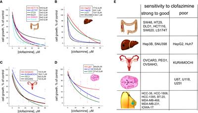
Wnt signaling plays key roles in oncogenic transformation and progression in a number of cancer types, including tumors in the breast, colon, ovaries, liver, and other tissues. Despite this importance, no therapy targeting the Wnt pathway currently exists. We have previously shown that the anti-mycobacterium drug clofazimine is a specific inhibitor of Wnt signaling and cell proliferation in triple-negative breast cancer (TNBC). Here, we expand the applicability of clofazimine to a set of other Wnt-dependent cancers. Using a panel of cell lines from hepatocellular carcinoma, glioblastoma, as well as colorectal and ovarian cancer, we show that the efficacy of clofazimine against a given cancer type correlates with the basal levels of Wnt pathway activation and the ability of the drug to inhibit Wnt signaling in it, being further influenced by the cancer mutational spectrum. Our study establishes the basis for patient stratification in the future clinical trials of clofazimine and may ultimately contribute to the establishment of the Wnt pathway-targeted therapy against a diverse set of cancer types relying on the oncogenic Wnt signaling.

View on the web

Neutrophils as Orchestrators in Tumor Development and Metastasis Formation

pay.thelady61 shared this article with you from Inoreader

581457_Thumb_400.jpg

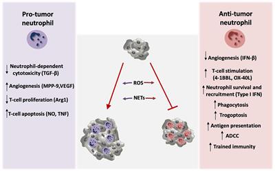
Several lines of clinical and experimental evidence suggest that immune cell plasticity is a central player in tumorigenesis, tumor progression, and metastasis formation. Neutrophils are able to promote or inhibit tumor growth. Through their interaction with tumor cells or their crosstalk with other immune cell subsets in the tumor microenvironment, they modulate tumor cell survival. Here, we summarize current knowledge with regards to the mechanisms that underlie neutrophil–mediated effects on tumor estab lishment and metastasis development. We also discuss the tumor-mediated effects on granulopoiesis and neutrophil precursors in the bone marrow and the involvement of neutrophils in anti-tumor therapeutic modalities.

View on the web

Qualitative MR Imaging Features for the Differentiation of Glioblastoma and Brain Metastases

pay.thelady61 shared this article with you from Inoreader

581037_Thumb_400.jpg

Objectives

To identify qualitative VASARI (Visually AcceSIble Rembrandt Images) Magnetic Resonance (MR) Imaging features for differentiation of glioblastoma (GBM) and brain metastasis (BM) of different primary tumors.

Materials and Methods

T1-weighted pre- and post-contrast, T2-weighted, and T2-weighted, fluid attenuated inversion recovery (FLAIR) MR images of a total of 239 lesions from 109 patients with either GBM or BM (breast cancer, non-small cell (NSCLC) adenocarcinoma, NSCLC squamous cell carcinoma, small-cell lung cancer (SCLC)) were included. A set of adapted, qualitative VASARI MR features describing tumor appearance and location was scored (binary; 1 = presence of feature, 0 = absence of feature). Exploratory data analysis was performed on binary scores using a combination of descriptive statistics (proportions with 95% binomial confidence intervals), unsupervised methods and supervised methods including multivariate feature ranking using either repeated fitting or recursive feature elimination with Support Vector Machines (SVMs).

Results

GBMs were found to involve all lobes of the cerebrum with a fronto-occipital gradient, often affected the corpus callosum (32.4%, 95% CI 19.1–49.2), and showed a strong preference for the right hemisphere (79.4%, 95% CI 63.2–89.7). BMs occurred most frequently in the frontal lobe (35.1%, 95% CI 28.9–41.9) and cerebellum (28.3%, 95% CI 22.6–34.8). The appearance of GBMs was characterized by preference for well-defined non-enha ncing tumor margin (100%, 89.8–100), ependymal extension (52.9%, 36.7–68.5) and substantially less enhancing foci than BMs (44.1%, 28.9–60.6 vs. 75.1%, 68.8–80.5). Unsupervised and supervised analyses showed that GBMs are distinctively different from BMs and that this difference is driven by definition of non-enhancing tumor margin, ependymal extension and features describing laterality. Differentiation of histological subtypes of BMs was driven by the presence of well-defined enhancing and non-enhancing tumor margins and localization in the vision center. SVM models with optimal hyperparameters led to weighted F1-score of 0.865 for differentiation of GBMs from BMs and weighted F1-score of 0.326 for differentiation of BM subtypes.

Conclusion

VASARI MR imaging features related to definition of non-enhancing margin, ependymal extension, and tumor localization may serve as potential imaging biomarkers to differentiate GBMs from BMs.

View on the web

A Nomogram Based on Serum Biomarkers and Clinical Characteristics to Predict Survival in Patients With Non-Metastatic Nasopharyngeal Carcinoma

pay.thelady61 shared this article with you from Inoreader

594363_Thumb_400.jpg

Objective

This study focused on developing an effective nomogram for improving prognostication for patients with primary nasopharyngeal carcinoma (NPC) restaged according to the eighth edition of the AJCC/UICC TNM staging system.

Methods

Based on data of 5,903 patients with non-metastatic NPC (primary cohort), we used Cox regression analysis to identify survival risk factors and created a nomogram. We used the nomogram to predict overall survival (OS), distant metastasis-free survival (DMFS) and disease- free survival (DFS) in the primary and independent validation (3,437 patients) cohorts. Moreover, we compared the prognostic accuracy between the 8th TNM system and the nomogram.

Results

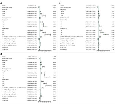
The nomogram included gender, age, T stage, N stage, Epstein–Barr virus DNA, hemoglobin, C-reactive protein, lactate dehydrogenase, and radiotherapy with/without induction or concurrent chemotherapy. In the prediction of OS, DMFS and DFS, the nomogram had significantly higher concordance index (C-index) and area under ROC curve (AUC) than the TNM system alone. Calibration curves demonstrated satisfactory agreements between nomogram-predicted and observed survival. The stratification in different groups permitted remarkable differentiation among Kaplan–Meier curves for OS, DMFS, and DFS.

Conclusion

The nomogram led to a more precise prognostic prediction for NPC patients in comparison with the 8th TNM system. Therefore, it could facilitate individualized and personalized patients' cou nseling and care.

View on the web

Macrophages in Osteosarcoma Immune Microenvironment: Implications for Immunotherapy

pay.thelady61 shared this article with you from Inoreader

586580_Thumb_400.jpg

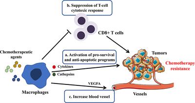
Osteosarcoma is a malignant primary bone tumor commonly occurring in children and adolescents. The treatment of local osteosarcoma is mainly based on surgical resection and chemotherapy, whereas the improvement of overall survival remains stagnant, especially in recurrent or metastatic cases. Tumor microenvironment (TME) is closely related to the occurrence and development of tumors, and macrophages are among the most abundant immune cells in the TME. Due to their vital roles in tumor progression, macrophages have gained increasing attention as the new target of tumor immunotherapy. In this review, we present a brief overview of macrophages in the TME and highlight the clinical significance of macrophages and their roles in the initiation and progression of osteosarcoma. Finally, we summarize the therapeutic approaches targeting macrophage, which represent a promising strategy in osteosarcoma therapies.

View on the web

Stereotactic Body Radiation Therapy for Extracranial Metastases From Primary Ovarian and Uterine Cancer

pay.thelady61 shared this article with you from Inoreader

572564_Thumb_400.jpg

Purpose

Single extracranial metastases from ovarian and uterine malignancies have historically been treated with surgery or conventional radiation. We report mature local control (LC), overall survival (OS), progression free survival (PFS), and toxicity for patients who completed 5-fraction stereotactic body radiation therapy (SBRT).

Methods

Patients with biopsy-proven, single extracranial metastases from primary ovarian and uterine malignancies treated with 5-fraction SBRT were included. Patients were stratified based on tumor volume (small < 50 cc or large ≥ 50 cc) and dose (low dose < 35 Gy or high ≥ 35 Gy). Kaplan–Meier method was used to estimate LC, OS, and PFS.

Results

Between July 2007 and July 2012, 20 patients underwent SBRT to a single extracranial metastasis. Primary site was divided evenly between ovarian and uterine (n = 10 each). Metastases involved the liver (30%), abdominal lymph nodes (25%), lung (20%), pelvic lymph nodes (10%), spine (10%), and extremity (5%). The median gross tumor volume (GTV) was 42.5 cc (range, 5–273 cc) and the median dose to the GTV was 35 Gy (range, 30–50 Gy). At a median follow-up of 56 months, the 5-year LC and OS estimates were 73 and 46%. When stratified by tumor volume, the 5-year LC and OS for small tumors were significantly better at 100% (p < 0.01) and 65% (p < 0.02). When stratified by dose, the 5-year LC was 87.5% with high dose and 53.6% with low dose (p = 0.035). The 5-year PFS for the entire cohort was 20%. Four patients with small metastases who had complete response remained disease free at study completion and were considered cured (median PFS > 10 years). Treatment was generally well tolerated, and only one patient experienced a late grade III musculos keletal SBRT related toxicity.

Conclusions

SBRT is a versatile, well-tolerated, and effective treatment option for single extracranial metastases from ovarian and uterine primary tumors. 35 Gy in five fractions appears to be a practical minimum effective dose. Four patients with small metastases were disease free at the study completion and considered cured. However, patients with larger metastases (≥50 cc) may require higher SBRT dosing or alternative treatments.

View on the web

Types of Mastectomies and Immediate Reconstructions for Ipsilateral Breast Local Recurrences

pay.thelady61 shared this article with you from Inoreader

567298_Thumb_400.jpg

Purpose: Ipsilateral-breast-local-recurrence (ILBLR) is a rare event with little data on immediate-breast-reconstruction (IBR). We report post-operative results of different types of mastectomy for ILBLR with or without IBR performed during a period of 40 months in order to analyze post-operative complications as main objective.

Methods: We analyzed mastectomies performed for ILBLR after initial breast conservative treatment from January 2016 to April 2019. The characteristics of patients, surgery, com plication rate, postoperative hospitalization have been determined.

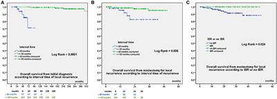
Results: Of the 207 mastectomies, 32.8% had an IBR: 31 nipple-sparing-mastectomy (NSM) and 37 skin-sparing-mastectomy (SSM) with 37 latissimus-dorsi-flap (LDF) IBR and 31 implant-IBR. Few reconstruction was performed for patients with body-mass-index ≥30 (OR = 0.214), infiltrating ductal carcinomas (OR = 0.272) and ASA-3 patients (OR = 0.254). In multivariate analysis, LDF-IBR was more often realized for NSM and for patients with BMI ≥25. The overall complication rate was 37.4%: 45.6 and 33.1% with and without IBR, respectively (p = 0.056). In multivariate analysis, BMI ≥25 (OR 2.02, p = 0.023), IBR (OR 1.9, p = 0.046) and tobacco (OR 2.17, p = 0.055) were correlated with higher risk of complications. There was no difference for Grade 2–3 complications rates for IBR and no IBR, respectively (14.7%: 10/68 and 9.3%: 13/139). In multivariate analysis, overall survival from date of mastectomy for local rec urrence was significantly associated with interval time to local recurrence (OR 6.981).

Conclusion: Salvage mastectomy and IBR is a good choice for ILBLR, particularly using flap reconstruction. NSM can be considered as a good option in selected patients for ILBLR for NSM and/or LDFR.

View on the web

Cepharanthine induces the proliferation of human dermal papilla cells and stimulates VEGF expression through increased intracellular calcium mobilization and HIF activation

pay.thelady61 shared this article with you from Inoreader

Summary

Background

Cepharanthine (CEP), which is extracted from Stephania cephalantha, is commonly prescribed to treat alopecia areata; however, the scientific evidence for its efficacy is limited.

Objective

To investigate the effect of CEP and its structural analogues on human hair growth in vitro.

Methods

The effects of CEP and three structural analogues of CEP on the proliferation of human dermal papilla cells (hDPCs) and human outer root sheath cells (hORSCs) were investigated. Their effects on vascular endothelial growth factor (VEGF) expression were also assessed by real‐time PCR. Finally, the activation of pathways leading to VEGF expression, such as intracellular Ca2+ mobilization and hypoxia‐inducible factor (HIF) expression, was characterized.

Results

CEP and two structural analogues of CEP significantly stimulated the growth of hDPCs but not hORSCs. Moreover, CEP and all three structural analogues significantly induced the expression of VEGF in hDPCs. CEP increased the intracellular Ca2+ concentration in hDPCs. CEP also increased the expression of HIF‐1α and HIF‐2α and induced the expression of HIF‐responsive genes in hDPCs, even under normoxia.

Conclusions

These results suggest that CEP and its structural analogues have the potential to restore hair growth by promoting the proliferation of hDPCs and increasing their expression of VEGF.

View on the web

The Role of Hippocampal Neurogenesis in ANT-DBS for LiCl-Pilocarpine-Induced Epileptic Rats

pay.thelady61 shared this article with you from Inoreader

?image=000509314-1.jpg

Purpose: Abnormal neurogenesis in the hippocampus after status epilepticus (SE) has been suggested as a key pathogeny of temporal lobe epilepsy. This study aimed to investigate the effect of deep brain stimulation of the anterior thalamic nucleus (ANT-DBS) on hippocampal neurogenesis in LiCl-pilocarpine-induced epileptic rats and to analyze its relationship with postoperative spontaneous recurrent seizures (SRS) and anxiety. Method: SE was induced by a syst emic LiCl-pilocarpine injection in adult male rats. Rats in the DBS group underwent ANT-DBS immediately after successful SE induction. SRS was only recorded during the chronic stage. An elevated plus maze was used to evaluate the level of anxiety in rats 7, 28, and 60 days after SE onset. After the elevated plus-maze experiment, rats were sacrificed under anesthesia in order to evaluate hippocampal neurogenesis. Doublecortin (DCX) was used as a marker for neurogenesis. Results: During the chronic stage, SRS in rats in the DBS group were significantly decreased. The level of anxiety was increased significantly in rats in the DBS group 28 days after SE, while no significant differences in anxiety levels were found 7 and 60 days after SE. The number of DCX-positive cells in the hippocampus was significantly increased 7 days after SE and was significantly decreased 60 days after SE in all rats in which SE was induced. However, the number of DCX-positive cells in the DBS gr oup was significantly lower than that in the other groups 28 days after SE. Conclusions: ANT-DBS may suppress SRS and increase the postoperative anxiety of epileptic rats by influencing hippocampal neurogenesis.
Stereotact Funct Neurosurg
View on the web

Efficacy of antimicrobial photodynamic therapy with Rose Bengal and blue light against cariogenic bacteria

pay.thelady61 shared this article with you from Inoreader

1-s2.0-S0003996920X00112-cov150h.gif

Publication date: Available online 9 December 2020

Source: Archives of Oral Biology

Author(s): Marina Hirose, Yasuo Yoshida, Kouichiro Horii, Yoshiaki Hasegawa, Yasuyuki Shibuya

View on the web

Antimicrobial peptides for the prevention and treatment of dental caries

pay.thelady61 shared this article with you from Inoreader

1-s2.0-S0003996920X00112-cov150h.gif

Publication date: Available online 9 December 2020

Source: Archives of Oral Biology

Author(s): John Yun Niu, Iris Xiaoxue Yin, William Ka Kei Wu, Quan-Li Li, May Lei Mei, Chu Chun Hung

View on the web

Αναζήτηση αυτού του ιστολογίου

! # Ola via Alexandros G.Sfakianakis on Inoreader