Objectives
To identify qualitative VASARI (Visually AcceSIble Rembrandt Images) Magnetic Resonance (MR) Imaging features for differentiation of glioblastoma (GBM) and brain metastasis (BM) of different primary tumors.
Materials and Methods
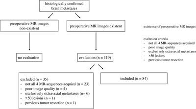
T1-weighted pre- and post-contrast, T2-weighted, and T2-weighted, fluid attenuated inversion recovery (FLAIR) MR images of a total of 239 lesions from 109 patients with either GBM or BM (breast cancer, non-small cell (NSCLC) adenocarcinoma, NSCLC squamous cell carcinoma, small-cell lung cancer (SCLC)) were included. A set of adapted, qualitative VASARI MR features describing tumor appearance and location was scored (binary; 1 = presence of feature, 0 = absence of feature). Exploratory data analysis was performed on binary scores using a combination of descriptive statistics (proportions with 95% binomial confidence intervals), unsupervised methods and supervised methods including multivariate feature ranking using either repeated fitting or recursive feature elimination with Support Vector Machines (SVMs).
Results
GBMs were found to involve all lobes of the cerebrum with a fronto-occipital gradient, often affected the corpus callosum (32.4%, 95% CI 19.1–49.2), and showed a strong preference for the right hemisphere (79.4%, 95% CI 63.2–89.7). BMs occurred most frequently in the frontal lobe (35.1%, 95% CI 28.9–41.9) and cerebellum (28.3%, 95% CI 22.6–34.8). The appearance of GBMs was characterized by preference for well-defined non-enha ncing tumor margin (100%, 89.8–100), ependymal extension (52.9%, 36.7–68.5) and substantially less enhancing foci than BMs (44.1%, 28.9–60.6 vs. 75.1%, 68.8–80.5). Unsupervised and supervised analyses showed that GBMs are distinctively different from BMs and that this difference is driven by definition of non-enhancing tumor margin, ependymal extension and features describing laterality. Differentiation of histological subtypes of BMs was driven by the presence of well-defined enhancing and non-enhancing tumor margins and localization in the vision center. SVM models with optimal hyperparameters led to weighted F1-score of 0.865 for differentiation of GBMs from BMs and weighted F1-score of 0.326 for differentiation of BM subtypes.
Conclusion
VASARI MR imaging features related to definition of non-enhancing margin, ependymal extension, and tumor localization may serve as potential imaging biomarkers to differentiate GBMs from BMs.

Δεν υπάρχουν σχόλια:
Δημοσίευση σχολίου
Medicine by Alexandros G. Sfakianakis,Anapafseos 5 Agios Nikolaos 72100 Crete Greece,00302841026182,00306932607174,alsfakia@gmail.com,